Tissues including membranes and glands: types
Definition
A tissue is a group of cells the Usally have a common original in an embryo and function together to carry out specialized activities.
Types of tissues
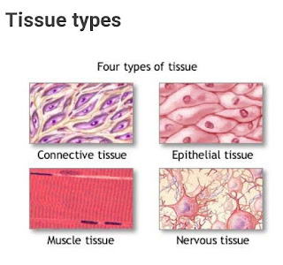
The tissues of the consist of large numbers of cells and they are classified according to the size, shape and functions of these cells. There are four main types of tissue that each have subdivisions . They are:
¹. Epithelial tissue
². Connective tissue
³. Muscular tissue
4. Nervous tissue
1. Epithelial Tissue
It covers body surface and lines hollow organs. Body cavities and ducts. It is allows the body interact with internal and y environment. Epithelial tissue consists of cells arranged in continuous sheets in single or multiple layers.
Structure of Epithelial Tissue:
Apical surface of an Epithelial cell faces the body surface, body cavity, lumen of an internal organ recieves cell secretion. The lateral surfaces of an Epithelial cell facing the adjacent cells on either side may contain tight junctions, desmosomes . The basal surface of an Epithelial cell is opposite to the apical layer refers to most superficial layer and basal layer is deepest layer . Basement is a thin extracellular layer commonly consists of two layers:
A tissue is a group of cells the Usally have a common original in an embryo and function together to carry out specialized activities.
Types of tissues
The tissues of the consist of large numbers of cells and they are classified according to the size, shape and functions of these cells. There are four main types of tissue that each have subdivisions . They are:
¹. Epithelial tissue
². Connective tissue
³. Muscular tissue
4. Nervous tissue

1. Epithelial Tissue
It covers body surface and lines hollow organs. Body cavities and ducts. It is allows the body interact with internal and y environment. Epithelial tissue consists of cells arranged in continuous sheets in single or multiple layers.
Structure of Epithelial Tissue:
Apical surface of an Epithelial cell faces the body surface, body cavity, lumen of an internal organ recieves cell secretion. The lateral surfaces of an Epithelial cell facing the adjacent cells on either side may contain tight junctions, desmosomes . The basal surface of an Epithelial cell is opposite to the apical layer refers to most superficial layer and basal layer is deepest layer . Basement is a thin extracellular layer commonly consists of two layers:
1. Basal lamina - Basal lamina contains proteins surch as glycoproteins, proteoglycons, collagen.
2. Reticular lamina contains collagen called Fibroblasts.
Functions:
It has three major functions. They serve as:
* Selective barriers that tranfer substances in and out of the body.
* Secretary surface that release products produced by the cells.
* Protective surface.
* Basement membranes have functions during wound healing and participate in filteretion of blood in kidneys.
* Epithelial Tissue play many different roles in the body such as protection, filteretion, secretion, absorption and excretion.
* In addition epithelial tissue combine with nervous tissue to form social organs for smell, hearing vision and touch.
a. Simple Epithelium:
1. Simple squamous epithelium: It is a simple layered with centrally located Nucleolus and is flattened, oval or spherical in shape
Location: Cardiovascular system and lymphatic vessels, kidney.
Functions: Blood filteration in kidneys. Diffusion of O² into blood vessels of lungs.
2. Simple Cuboidal Epithelium: Simple layer of cube - shaped cells, centrally located nucleus
Location : Covers surface of ovary, secreting portion of some gland
Function : secretion and absorption.
3. Simple Columnar Epithelium (non-ciliated) : Single layer of columnar cells with oval nuclei near base of cells. It contains goblet cells.
Location : Lines of gastro intestinal tract, gall bladder.
Function : secretion and absorption
4. Pseudostratified Columnar Epithelium : It appears to have several layers. Pseudo stratified ciliated columnar epithelium contain cells extend to surface and secrete mucus.
5. Pseudo stratified non-ciliated columnar epithelium : it contains cells without cilia and lacks goblet cells.
Location : Upper respiratory tract, epididymis.
Function : Secretes mucus traps foreign particles, absorption and protection.
b. Stratified Epithelium :
1. Stratified squamous epithelium :
It consists of two or more layers of cells.
* Stratified squamous keratinized Epithelium develops tough layer of keratin in apical layer of cells and several layers deep to it.
*Stratified squamous non-keratinized Epithelium does not contain large amounts of keratin in apical layer and several layers deep and is constantly moistened by mucus from salivary and mucous glands.
Location : keratinized is seen in skin . Non-keratinized is seen in oesophagus, tongue vagina.
Function : Protection, water loss, defence.
2. Stratified Cuboidal Epithelium :
It consists of two or more layers of cells.
Location : ducts of adult sweat gland.
Function : Protection, secretion and absorption.
3. Stratified Columnar Epithelium :
Basal layers usually consist of shortened irregular- shaped cells. Apical layer has columnar cell.
Location : Lines part of male Urethra, conjunctiva of eye, esophageal gland.
Functions : Protection, secretion.
4. Transitional epithelium : When relaxed looks like stratified Cuboidal Epithelium, apical layer is large and umbrella-shaped . When stretched. Cell becomes flattened and multiple layers are seen .
Location: Lines urinary bladder, ureters and urethra.
Function : Allows urinary organs to stretch and maintain protective lining.
2. Connective Tissue
Connective tissues are one of the most abundant and widely distributed tissues in the body. Connective tissue consists of two basic elements extracellular matrix and cells. Extracellular matrix consists of protein fibers and ground substance the material between the cells and the fibers. The different types of cells involved in connective tissue are: Fibroblasts, fat , cells, macrophages, Leucocytes and mast cells.
Function : Protection , support, Immunity, Reservoir of energy.
Classification of Connective Tissue : Connective Tissue can be classified into two types :
1. Embryonic Connective Tissue : it is present primarily in the embryo, the developing human form fertilization through the first two months of pregnancy and in the fetus the developing human form the third month of pregnancy. It has mesenchyme and mucous connective tissue.
2. Mature Connective Tissue : it is divided into :
A. Loose connective tissue
B. Dense connective tissue
C. Cartilage
D. Bone tissue
E. Liquid connective tissue
A. Loose Connective Tissue :
These are loosely arranged between the cells. Types of loose connective tissues are :
Losse connective tissue
A. Areolar connective tissue
B. Adipose tissue
C. Reticular connective tissue
A. Areolar connective tissue:
This is most widely distributed connective tissue consisting of fibres arranged randomly and several kinds of cells embedded in semifiuid ground substance.
Function : structure, elasticity, support
B. Adipose tissue :
It has cells derived from Fibroblasts called adipocytes. Cells fill up with a single large triglyceride droplets, cytoplasm and nucleus are pushed to periphery of cell. These are located deep to skin , around heart and kidneys, yellow bone marrow.
Function : Reduced heat loss , protection energy reserve
C. Reticular connective tissue :
These are fine interlacing network of reticular fibers and reticulum cells. It is located in liver , spleen , nodes , around blood vessels and muscles .
Function : forms stroma of organ filters and removes worn out blood cells in spleen and microbes in lymph nodes.
B. Dense Connective Tissue :
It contains more fibers and fewer cells than loose connective tissue. These are of three types :
1. Dense Regular Connective Tissue:
It is arranged in parallel bundles . It has great tensile strength that resit pulling forces especially well in one direction.
2. Dense Irregular Connective Tissue :
It has fibers that are not arranged in parallel bundles ad in dense regular connective tissue.
3. Elastic Connective Tissue :
These are bundles of proteins found in extracellular matrix of connective tissue produced by Fibroblasts and smooth muscle cells in arteries.
C. Cartilage :
Cartilage is firmer than other connective tissues. The cells are called chondrocytes and are less numerous. They are embedded in matrix reinforced by collagen and elastic fibres. They are of three types:
1. Hyaline cartilage :
It is a smooth bluish white tissue. The chondrocytes are in small groups within cell nests and the matrix is solid and smooth. It consists of perichondrium .
* Hyaline cartilage provides flexibility, support and smooth surfaces for movement at joints.
* it is found at the ends of long bones that form joints.
* Forming the costal cartilages which ribs to sternum.
* Forming part of larynx, trachea and bronchi.
2. Fibro cartilage :
This consists of dense of dense masses of white collagen fibers in a matrix similar to that of hyaline cartilage with the cells widely dispersed. It is tough , slightly flexible, supporting tissue found in ligaments joints and bones .
* Between articulating surfaces of bones .
As pads between bodies of vertebrae.
3. Elastic cartilage :
This flexible tissue consists of yellow elastic lying in a solid matrix. The chondrocytes lie between the fibers . It provides support and maintains shape
D. Bone tissue:
bone cells are surrounded by a matrix of collagen fibres strengthened by inorganic salts especially calcium and phosphate. This provides bones with their characteristics strength and rigidity. These are classified as :
1. Compact Bone:
The basic unit of compact bone is ostean or haversian system. Each
Ostean has for parts.
* Lamellae :
These are concentric rings of extracellular matrix that consists of mineral salt which gives to bones hardness and strength.
* Lacunae :
Consists of osteocytes.
* Canaliculi :
It provides route for nutrient to reach osteocytes.
* Haversian canal :
Haversian canal contains blood vessels and nerves.
2. Spongy or cancellous bone :
Spongy bone lacks osteons .it consists of columns of trabeculae . Spance between trabeculae are filled with red bone marrow.
E. Liquid connective tissue:
* Blood tissue is connective tissue with a liquid extracellular matrix and formed elements blood plasma is a yellow fluid consisting of mostly water and dissolved substances nutrients, wastes hormones, ions formed elements present in blood plasma are RBC WBC , platelets etc.
* Lymph :
Is an extracellular fluid that flows in lymphatic vessels. The composition of lymph varies from one part of the body to another.
3. Muscular tissue :
Muscular tissue consists of elongated cells called muscle fibers. Muscle tissue is able to contract and relax providing movement within the body . Muscle contraction requires an adequate blood supply to provide sufficient O², Ca²+ and nutrients and to remove wast products . It is divided as three types.
1 . Skeletal Muscle :
It consists of striated and voluntary muscle fibres that are cylindrical and contain several nuclei 35 cm long.
2. Smooth muscle :
It is a nonstriated involuntary muscle. It consists of central spindle
Shaped nucleus.
3. Cardiac muscle:
It is found in wall of heart it is striated muscle consisting of Nucleolus and interconnected discs.
4. Nervous tissue
Nervous tissue is made up of different types of nervous cells, all of which have an axon the long stem like part of the cell that sends A.P. ( Action Potential) signals to next cell . Nervous tissue is composed of neurons or nerve cells which receive and transmit impulses. Nervous tissue is specialized to react to stimuli and to conduct impulses to various organs in the body which brings about a response to the stimulus.
Comments
Post a Comment